
Transperineal Pelvic Floor Ultrasound Scan
In this Health Topic
This is a scan of the pelvic floor using a hand-held probe or ‘transducer’ which is placed on the perineum (the area between the vagina and the anus). This enables images to be taken of the bladder, urethra (what urine flows through), vagina, cervix, uterus (womb), perineum, anal canal, and pelvic floor muscles. See images below.
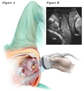
Figure A shows how the transducer is placed on the perineum. Figure B shows the pelvic floor organs: SP-pubic symphysis (pubic bone), U-urethra, BL-bladder, Ute-uterus, V-vagina, AC-anal canal, Rec-rectum (lowest part of the bowel).
You may be asked to empty your bladder just before the scan. For best results, it is better if you have also had a recent bowel movement.
The scan is performed by a trained health care professional. After undressing from the waist down, you will be asked to lie down on an examination chair or table and cover yourself with a sheet or drape. The lights will be dimmed to provide clearer visualization of the screen during the scan. You will then be asked to bend your knees and bring your heels as close to your buttocks as possible. After applying gel on the transducer and covering it with a powder free glove, more gel is applied on the transducer which will then be placed on the perineum. This might feel a bit cold, but the scan itself does not produce any major discomfort. You may feel the pressure of the probe. Sometimes it is necessary to part the labia (lips of the vagina) and the probe might be moved in different directions. During the scan, you will be asked to squeeze and bear down a few times to obtain precise information. The person doing the scan will explain exactly what you need to do. Sometimes it might be necessary to perform the scan with you in a standing position.
The scan will show pelvic floor problems such as prolapse of the bladder, uterus, rectum and bowel. In addition, it will show if there has been previous damage to the pelvic floor muscles and/or anal canal during childbirth. If you previously had ‘mesh implants’ or a sling inserted, these may also be seen on the scan. Other findings that can be seen include incomplete emptying of the bladder, proper functioning of the pelvic floor during muscle contraction, and complex problems of the back passage such as a prolapse of the vaginal back wall (rectocele) or rectal intussusception (when the rectum telescopes on itself). These findings will assist your doctor with planning appropriate treatment.